Medial knee pain that doesn't quite behave like a meniscus tear is one of those presentations that can send you down the wrong path if you're not thinking about the plica. It's an easy structure to overlook because it doesn't show up clearly on imaging the way a meniscus tear often does, and its symptoms can overlap so much with meniscal pathology that it gets missed or misdiagnosed fairly often.
What the Medial Plica Actually Is
The medial plica is a fold of synovial tissue left over from how the knee develops before birth. During early development the knee joint is divided into compartments by these synovial folds, and normally most of that tissue resorbs before birth, but remnants persist in a large portion of the population, with the medial plica being one of the more common ones to stick around. Sitting between the medial border of the patella and the medial femoral condyle, near the adductor tubercle region, it's usually harmless and asymptomatic.
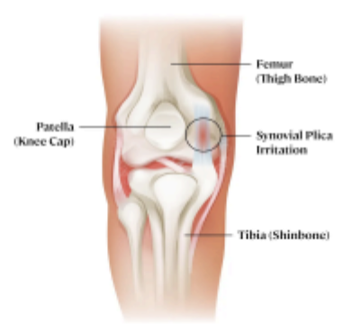
The problem starts when it becomes irritated, most often from repetitive knee movement, direct trauma, or overuse. Once irritated, the plica can thicken and lose its normal elasticity, and a thickened, fibrotic plica can start snapping or catching over the medial femoral condyle during knee movement. That repetitive friction can lead to synovitis and even chondral irritation over time, and separately, a tight or tethered plica can interfere with normal quadriceps mechanics, which adds another source of anterior or medial knee pain that isn't purely about local tissue irritation.
Why It Gets Confused With a Meniscus Tear
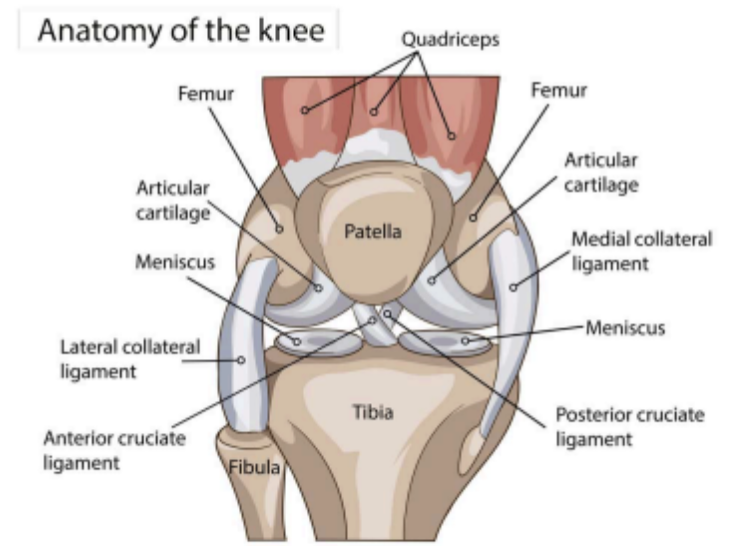
The overlap here is real, not just a case of poor clinical reasoning. Anatomically the medial plica sits right next to the medial meniscus, so when one structure is irritated it can quite literally rub against and aggravate the other. That means these two problems don't just look similar, they can actually feed into each other, an inflamed plica can aggravate a meniscus, and a meniscus injury can irritate a nearby plica in return.
Imaging doesn't always save you here either. Even MRI can struggle to clearly distinguish an inflamed or thickened plica from meniscal pathology, since both can present similarly on scans. Because of that, plica syndrome is still considered largely a clinical diagnosis, built from history and physical exam rather than imaging alone, and it remains one of the more commonly missed knee diagnoses for exactly that reason.
How to Differentiate Clinically
1. History and pain behaviour: Meniscus tears often relate to a clear twisting or loading mechanism, and pain tends to sit more at the joint line itself. Plica irritation more often
builds gradually with repetitive movement, or follows a direct knock to the front of the knee, and pain tends to sit slightly more anterior and medial, closer to the femoral condyle than the true joint line. Snapping, catching, or a sensation of something rolling under the skin with flexion and extension leans more toward plica than meniscus.
2. Palpation: For the plica, palpation involves rolling your fingers over the fold of tissue located between the medial border of the patella and the medial femoral condyle, near the adductor tubercle. Under your fingers it should feel like a distinct ribbon-like band of tissue rolling against the condyle beneath it, and reproducing mild pain or tenderness there, especially compared with the same spot on the uninvolved knee, supports the diagnosis. This is different from meniscal tenderness, which localises more precisely to the actual joint line rather than slightly proximal and anterior to it.
3. Special tests
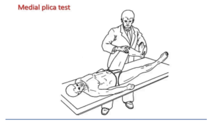
a. Mediopatellar plica test or Hughston's plica test: the knee is moved through flexion and extension while the examiner applies pressure over the plica region, feeling for a palpable snap or reproducing the patient's pain, which supports plica involvement.
b. McMurray's test and joint line tenderness remain the classic tools for meniscal pathology, looking for a click or pain specifically with rotation and compression through the joint line rather than the more medial, condyle level tenderness seen with plica.
c. The Thessaly test is another one worth using alongside joint line tenderness when you're leaning toward meniscal involvement, since it loads the joint through weight bearing rotation rather than passive positioning.
None of these tests are highly specific on their own, so a cluster of findings, history, palpation location, and test response together, is far more useful than relying on a single special test.
Other structures worth ruling out
1. Pes anserine bursitis or tendinopathy, tender lower and more posterior along the medial tibia rather than at the joint line or condyle.
2. Medial collateral ligament sprain, tender along the ligament's course with a valgus stress mechanism and positive valgus stress test.
3. Patellofemoral pain, more diffuse anterior knee pain related to loading through squatting, stairs, or prolonged sitting rather than a focal medial tender point.
How to Rehab It as a Physio
The encouraging thing here is that conservative management genuinely works for a lot of people, and it's generally the sensible starting point before anything more invasive is considered.
1. Settle the irritation first
Relative rest from the aggravating repetitive movements, activity modification, and anti-inflammatory strategies form the early stage. This isn't about full rest, it's about reducing the specific repetitive knee flexion and extension load that's keeping the plica irritated while still keeping generally active.
2. Restore normal quadriceps mechanics
Since a tight or tethered plica can alter normal quadriceps function, addressing quadriceps activation and control, particularly VMO timing and patellar tracking, helps reduce the abnormal tension being placed through the plica's synovial attachment. Soft tissue work around the
quadriceps and surrounding retinaculum can help here too, similar in principle to how we'd address any tissue that's become mechanically tethered.
3. Gradually reload through range
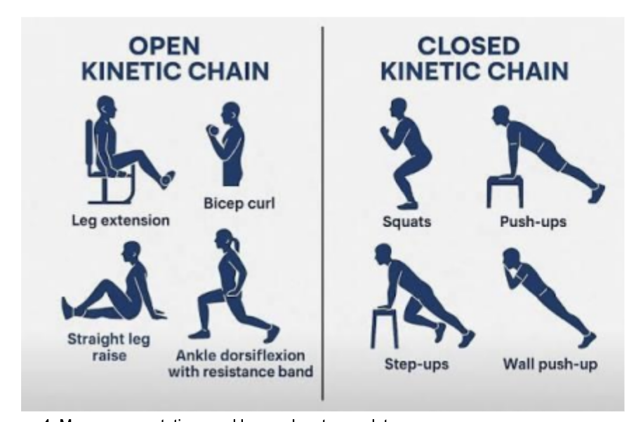
Once acute irritation has settled, progressively reintroducing full range knee flexion and extension under control helps desensitise the snapping or catching sensation. Closed chain strengthening, squats, step ups, and lunges progressed through range as tolerated, tends to work better than isolated open chain quad work alone, since it trains the knee to move well under real load rather than just building isolated strength.
4. Manage expectations and know when to escalate
Most people do improve with a structured conservative approach, but it's worth knowing that when conservative management and structured physiotherapy have genuinely been given a fair trial and symptoms aren't settling, arthroscopic resection of the plica is a well supported next step, and research comparing the two head to head has found better outcomes with surgery in patients who hadn't responded to structured physiotherapy first. That's not a reason to rush toward surgery, but it is a useful thing to communicate to a patient early, that conservative care is absolutely worth trying properly first, but it isn't the only option if things stall.


